What is Femoroacetabular Impingement of the Hip?
Femoroacetabular impingement (FAI) is a painful condition caused by irritation and inflammation of the soft tissues of the hip joint. This can occur because the femoral head (the ball of the hip) pinches up against the acetabulum (cup of the hip joint). This can be due to extra bone growth, on one or both of the bones that form the hip joint, giving the bones an irregular shape. When this happens the bones don’t fit together properly, and therefore rub against each other during movement. Over time this friction can damage the joint, tendon and/or the cartilage surrounding the joint (known as the labrum), causing pain, stiffness and limited activity.
If left untreated it may increase a person’s risk of developing chronic hip joint conditions such as osteoarthritis.
For this reason, timely and effective treatment is essential, for all stages of hip impingement. In this post, we will discuss hip impingement symptoms, causes, and effective treatment options.
Anatomy of the Hip Joint
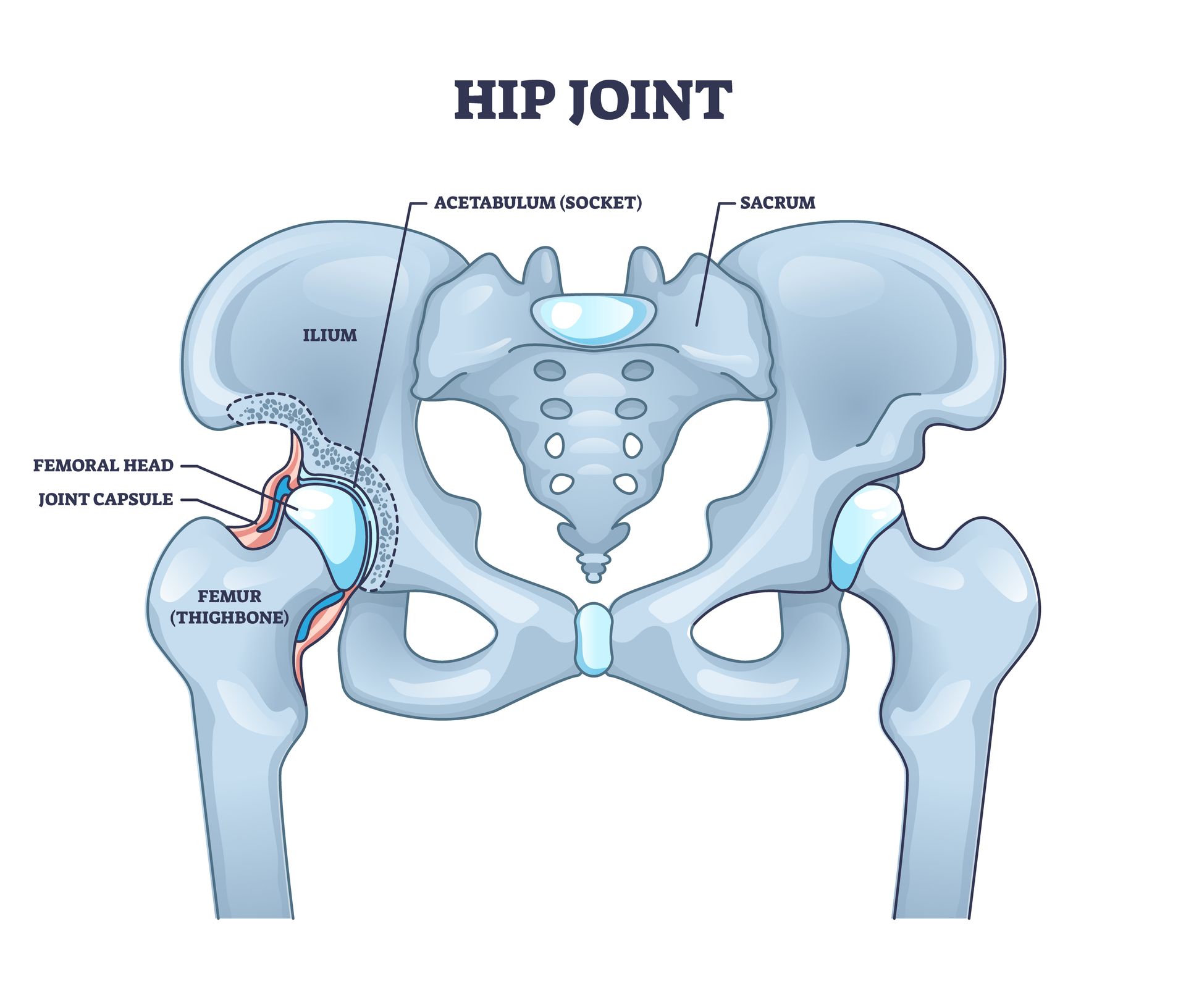
The hip joint is what’s called a ball and socket joint. The acetabulum in your pelvis comprises the “socket” portion and the femoral head, or the top portion of the thigh bone, creates the “ball” portion of the hip joint. It is one of the largest and most important joints in the body (see image below).
A thick soft tissue layer called articular cartilage covers the surface of the bones i.e. the ball and
the socket. This layer of cartilage creates a smooth, low friction surface that ensures the bones
glide easily over each other during movement.
There is an outer rim of cartilage (fibrocartilage) known as the labrum that surrounds the
acetabulum. The labrum adds extra stability to the joint by deepening the socket of the
acetabulum, and acts to seal off the joint fluid to help keep the joint lubricated. The labrum also
allows the ball and socket joint to operate smoothly during activity.
What Causes Femoroacetabular Impingement of the Hip?
The abnormalities associated with FAI are often present at birth. But they can develop later in life,
especially during the teenage years. When the hip bones are shaped abnormally, there is little that
can be done to prevent FAI.
There are three types of anatomical variation which can lead to femoroacetabular hip:
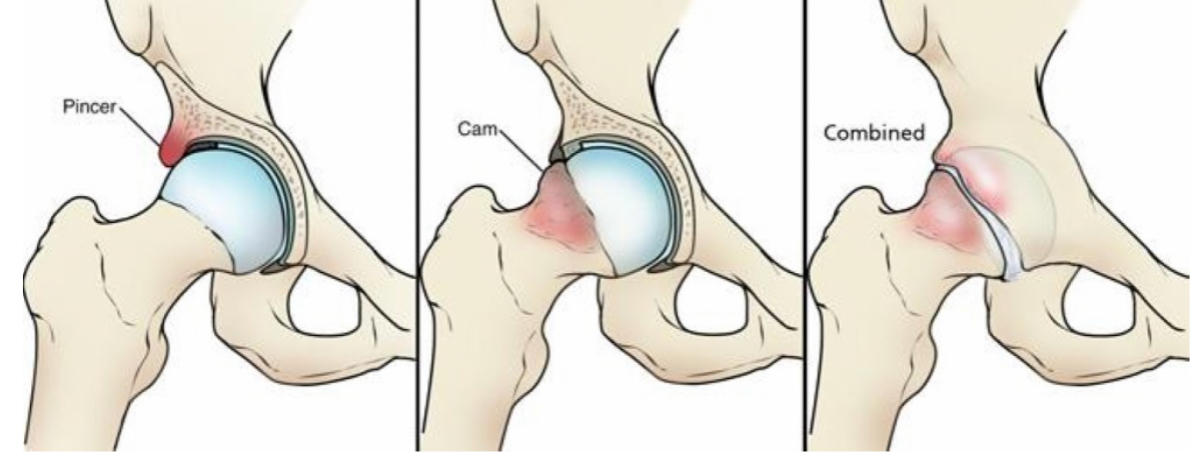
There are three types of anatomical variation which can lead to femoroacetabular hip impingement:
Pincer: This type of impingement is caused by extra bone growth which extends over the rim of the acetabulum in the hip socket. The labrum can become pinched by the prominent rim.
Cam: In this type of impingement, the ball has a more oval than round appearance. This means that the head cannot rotate smoothly in the acetabulum, creating friction when the ball hits the edge of the socket. This repeated friction can cause bony growth on the head of the joint, adding to further impingement.
Combined: This means that both the cam and pincer types of FAI are present.
When one, or both, of these lesions are present, they can cause pinching or impingement of the hip joint structures; mainly the labrum around the edge of the joint. As previously described, the labrum is a layer of cartilage which encircles the socket of the hip joint. It is susceptible to damage when repetitive impingement takes place. Over time this impingement of the hip joint can cause pain, especially with prolonged periods of sitting or during and after exercise.
Risk Factors Associated with FIA
Research suggests that the development of femoral acetabular impingement is mainly congenital; i.e. you are born with it. However, there are a number of risk factors associated with femoral acetabular impingement (FAI). These include;
- Hip muscle weakness especially the gluteal muscles
- Hypomobility (reduced range of movement) or hypermobility (excessive range of movement) of the hip joint
- Muscular stiffness/tightness in the muscles surrounding the hip
- Altered gait (walking) pattern
- Previous hip injury such as a fracture
- Pelvic posture
- Type of sporting activity carried out at a young age (adolescent years when growing). For example, it has been shown that those who participate in football at a young age have a predisposition to developing FAI secondary to a CAM lesion. Because active people tend to work the hip joint more vigorously, they may begin to experience pain earlier than those who are less active. However, exercise does not cause FAI.
What are the Symptoms of Femoroacetabular Impingement of the hip?
You can have hip impingement for a number of years and not know it, because it is often not painful in its early stages.
When hip impingement causes symptoms, the main symptoms are pain in the groin and outside of the hip. Often the pain is aggravated when turning, twisting, running or squatting and accompanied with hip stiffness i.e. decreased range of motion in the hip. The pain can be sharp with movement, but is also described as a more constant dull ache.
- The most common symptoms of FAI include:
- Moderate to marked hip or groin pain with certain hip movements or positions
- Pain can also be reported in the thigh, back or buttock
- Stiffness and reduced movement in the hip joint
- Clicking and/or catching sensation in the hip
- Locking or giving way
- Decreased ability to perform activities of daily living and sports
- Limping when walking or running
How do we Treat Femoroacetabular Impingement of the Hip?
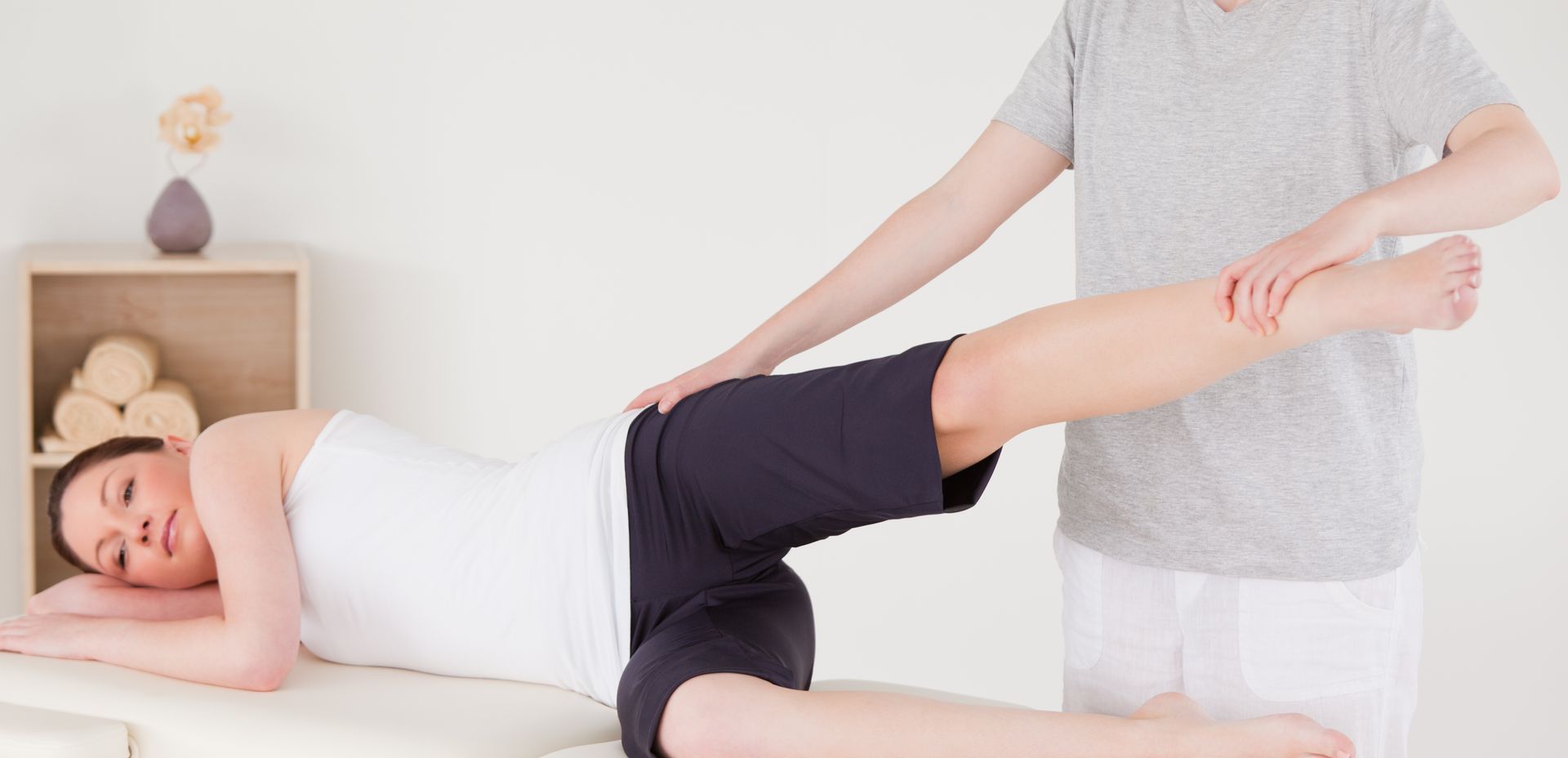
Femoroacetabular impingement normally responds extremely well to physiotherapy and activity modification.
While it is not possible to reverse the bone and joint changes that occur with FIA, there are a number of treatment options available to help reduce your pain and symptoms, and allow you to live a full and active life. The main treatment goals of physiotherapy for FIA are:
- Reduce your hip pain and joint inflammation.
- Increase your hip joint range of movement and muscle flexibility.
- Strengthen your lower limb muscles.
- Improve your balance, proprioception and agility.
- Optimise your functional movements and activity level, e.g. walking, squatting, climbing stairs.
- Physiotherapy Treatment for Hip Femoroacetabular Impingement
Your physiotherapist will provide a specific, tailored, treatment and rehabilitation programme, which may include:
- Relative Rest – Advice activity modification and rest, to allow your pain to settle and minimise the chance of irritating your symptoms further. Prolonged sitting in low chairs and sitting cross legged should be avoided.
- Patient Education – Understanding the cause of your symptoms is an important part of your treatment process. It can give you peace of mind and also help you to take a more active role in your rehabilitation.
- Pain Management – Painkillers and non-steroidal anti-inflammatory medication can often be useful in the early stages of management (any medication needs to be approved by a pharmacist or doctor).
- Manual (hands on) Therapy – Your physiotherapist may use manual therapy such as joint mobilisations or soft tissue techniques, to release the joints, muscles and soft tissues. This will help improve joint movement and reduce muscle stiffness or soreness.
- Range of Movement Exercise – Your physiotherapist may teach you some stretching exercises for your hip joint and surrounding muscles, to help restore normal movement of your hip.
- Hip Strengthening Exercises – Your physiotherapist will design a progressive resistance program to ensure all your lower limb muscle groups are strong. It will specifically target your deep gluteal muscles, which will help to stabilise your hip joint.
- Proprioception and Balance Exercises – Damage to your joints and soft tissues, can have a negative affect on your balance. These exercises will help your brain to reconnect with your hip, and give you a better sense of balance during movement and activities.
- Biomechanical and movement re-education – Lower limb joint injuries can alter your movements, especially walking and running. Your physiotherapist may need to re- teach you how to move correctly.
- Functional and Sports Specific Training – Once your pain, movement and strength have improved, your physio will work on specific, functional training to help you safely resume more demanding activities and sports.
Chelmsford Physio
Riverside Leisure Centre, Victoria Rd, Chelmsford CM1 1FG